Book Appoinment
Call Now For Make an Appointment
022-25887210
Q1)what is Echocardiography?
A test called echocardiography uses sound waves to create live images of your heart. An echocardiogram is the name of the image. Your doctor can keep track of how your heart and its valves are working with this test.
They may be able to obtain information about the size of the heart, for example, if the chamber size, dilation, or thickness change blood clots in the heart chambers fluid in the sac around the heartproblems with the heart's pumping or relaxing function issues with the aorta, the main artery that connects the body to the heart problems with the heart's pumping or relaxing functions problems with the function of heart valves pressure in the heart.Especially after a heart attack, an echocardiogram is essential for determining the health of the heart muscle. Additionally, it may reveal heart defects or other irregularities in unborn children.
Getting an echocardiogram is painless. With certain types of echocardiograms or when contrast is used for the echocardiogram, there are only very rare risks.here are several different types of echocardiograms.
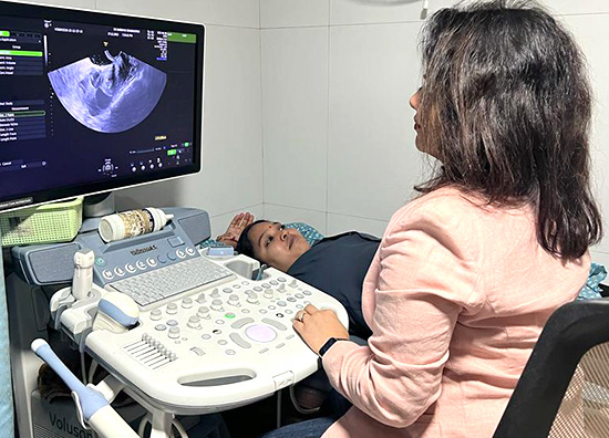
Transthoracic echocardiography
The most prevalent form of echocardiography is this one.You will have a transducer placed over your heart on your chest. Ultrasound waves travel through your chest and toward your heart through the transducer. As the sound waves return to the transducer, a computer interprets them. The live images that are displayed on a monitor are the result of this.A health specialist for the Top Echocardiography Testing Clinic in Vartak Nagar, Thane will follow guidelines for collecting different types of images and information.The diagnostic test known as 2D Echocardiography or 2D Echo is used to identify heart disease. It is a method for taking images of the heart using ultrasound. In fact, it provides a cross-sectional view of the heart, highlighting its chambers, valves, and major blood vessels. It is determined from the image how your heart looks and how its muscles work.. The purpose of the echocardiogram is to assess the structure and function of the heart. The information provided by the results will assist the healthcare provider in making a heart-related diagnosis.Echocardiograms can be repeated over time to keep an eye on how the heart is working. The results can help figure out if the previous treatment was effective and if it needs to be changed
Transesophageal echocardiogram
The heart's structure and function are examined with an echocardiogram during a transesophageal echocardiogram (TEE). A transducer, similar to a microphone, emits ultrasonic sound waves during the procedure. The ultrasonic sound waves travel through the skin and other body tissues to the heart tissues when the transducer is positioned at particular angles.The transducer is positioned on the chest's surface during a traditional echocardiogram.echocardiogram. A transducer-equipped probe is inserted down the esophagus for a transesophageal echocardiogram.Because the sound waves don't have to go through skin, muscle, or bone, this makes it easier to see the heart.
Contrast echocardiography
Contrast echo can help confirm an atrial septal defect (ASD) diagnosis.ASD). Agitating saline or synthetic contrast create microbubbles. When injected intravenously, these are very reflective and can cause opacification in the echo window. Before being absorbed and trapped by the pulmonary capillaries, they are typically seen on the right side of the heart and do not travel to the left.. The contrast created by the bubbles allows a left-to-right shunt to be seen as a jet "interrupting" the opacification of the right atrium.A right-to-left shunt, on the other hand, theoretically carries the possibility of systemic air embolism. When the transducer is positioned on the chest wall, being overweight or having certain lung diseases can cause images of the heart to be distorted. Mitral valve disorders, blood clots or masses inside the heart, and a tear in the lining of the heart are all better seen with TEE. the aorta, and the structure and function of artificial heart valves.
- M-mode echocardiography
- Doppler echocardiography.
- Color Doppler
- 2-D (two-dimensional) echocardiography
- 3-D (three-dimensional) echocardiography
Stress echocardiogram
Some heart problems — particularly those involving the arteries that supply blood to your heart muscle (coronary arteries) — occur only during physical activity. Your doctor might recommend a stress echocardiogram to check for coronary artery problems. However, an echocardiogram can't provide information about any blockages in the heart's arteries.
In a stress echocardiogramMUltrasound images of your heart are taken before and immediately after you walk on a treadmill or ride a stationary bike.If you're unable to exercise, you may get an injection of a medication to make your heart pump as hard as if you were exercising.Looking for the Top Echocardiography Testing Clinic in Vartak Nagar, Thane